Anne Seccombe
“We propose that in utero exposure to excessive levels of steroids such as estrogen has a long-term effect on the ability of the thymus to produce regulatory T cells. In female offspring this can lead to PCOS.”
Researchers from Binghamton University in New York published an article on 18 May 2009 in the Journal of Reproductive Biology and Endocrinology hypothesizing a new cause for PCOS.
Essentially they suggest, and their study supports, the theory that exposure of a foetus to high levels of steroids (such as eostrogen, testosterone or corticosteroids) whilst it is in utero may cause a cascade of auto-immune dysfunctions involving the thymus gland and it’s production of regulatory T-cells, eventually resulting in polycystic ovarian syndrome if the child is female. Sources of steroid exposure are likely to be from dietary phytoestrogens in the case of oestrogen or severe maternal stress whilst pregnant in the case of corticosteroids.
In the study, mice were injected with oestrogen at between 5 & 7 days old. This is known to cause anovulation and follicular cysts, a disease model that bears a similarity to PCOS in humans.
It is generally believed (but not proven) that this response is due to the hypothalamus responding to the excess oestrogen and a disruption in the GnRH delivery system.
This group of researchers, however, had a new idea which they sought to prove, namely that the anovulation and follicular cysts may be part of an immune response to the oestrogen and involving the thymus gland and it’s production of a specialised type of white blood cells called T cells and which help to fight infection or potential infection and also have a regulatory effect. High levels of oestrogen appear to prevent the thymus gland from producing fully developed regulatory T-cells. The absence of T-regulatory cells is known to cause a number of auto-immune diseases.
The researchers found that mice who had their thymus gland removed when they were 3 days old, prior to the injection of oestrogen, they did not develop follicular cysts and anovulation. When these mice later received an infusion from older mice who had been exposed to oestrogen, they found that the disease was transferable in a large percentage of the mice through the lymphocytes. Thus it would appear that the absence of regulatory T-cells is a prerequisite for the formation of follicular cysts.
The researchers concluded that exposure in utero to steroid hormones such as oestrogen or testosterone are unlikely to be derived from the mothers’ own hormones as these are usually only present in nanogram levels, whereas milligram levels are required to produce these effects. Therefore, the researchers hypothesize that it is more likely that these levels are achieved through the ingestion of phytoestrogens from the mothers’ diet, such as are found in soy and flaxseed products. Although it wasn’t covered in depth in the present study, the researchers highlighted that severe stress in pregnancy would raise levels of corticosteroids which are also known to negatively affect the spleen and thymus development of a foetus and the subsequent production of lymphocytes/thymocytes respectively.

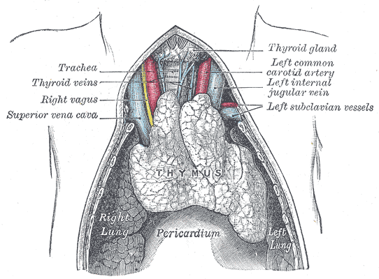
The thymus gland is located just behind the top of the sternum or breastbone, below the thyroid gland and acts as a nursery where lymphocyte precursors which are produced in the bone marrow are taken out of the blood into the thymus gland, where they mature into thymocytes and eventually T-cells. Once they are mature, the T-cells are released back out into the blood stream where they have a complex function in regulating the immune system.
The thymus gland is an interesting one, in that it is most active during childhood developing to it’s peak until puberty, where it stops developing and starts to atrophy. When a child is born, the thymus gland is pinkish-grey in colour and lobulated (it has a bumpy surface, somewhat like the brain). It weighs about 15 grams at birth and is about 5 cm long, 4 cm wide and 6 mm deep. It is separated into two lobes, so it looks something like a butterfly drawn by a 2 year old. By the time a child reaches puberty the thymus gland has grown to about 35 grams in weight and has developed all the T-lymphocytes it will need to last a lifetime. By the time an adult reaches their seventies, the gland has degenerated into fatty islands which are almost indistinguishable from the surrounding tissue and have turned yellow in colour. It will now weigh only about 5 grams; a third of it’s weight at birth, and less than 15 % of it’s peak weight at puberty.
More Information:
Chapman JC et al, The estrogen-injected female mouse: new insight into the etiology of PCOS, Reprod Biol Endocrinol. 2009 May 18;7(1):47
Interesting… It might also help to explain why women with PCOS also tend to have higher instances of inflammatory conditions.